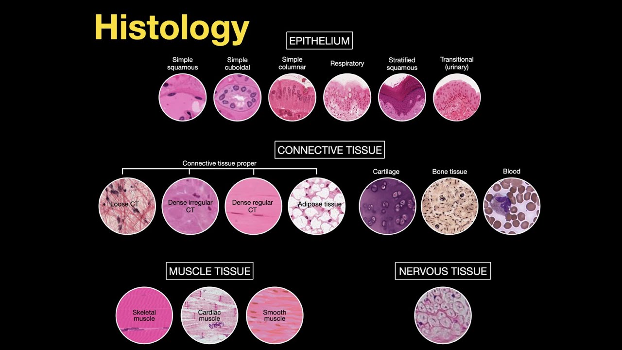
Professor Dave here, let’s check out some bones. We were first introduced to bones when we learned about connective tissue, as bone falls into this category, but now it’s time to get much more specific about the structure of bone, and take a look at all the types of bones found in the body. Before diving into the bones that we are familiar with, let’s start with the bits of skeletal cartilage that can be found in various regions of the skeleton.
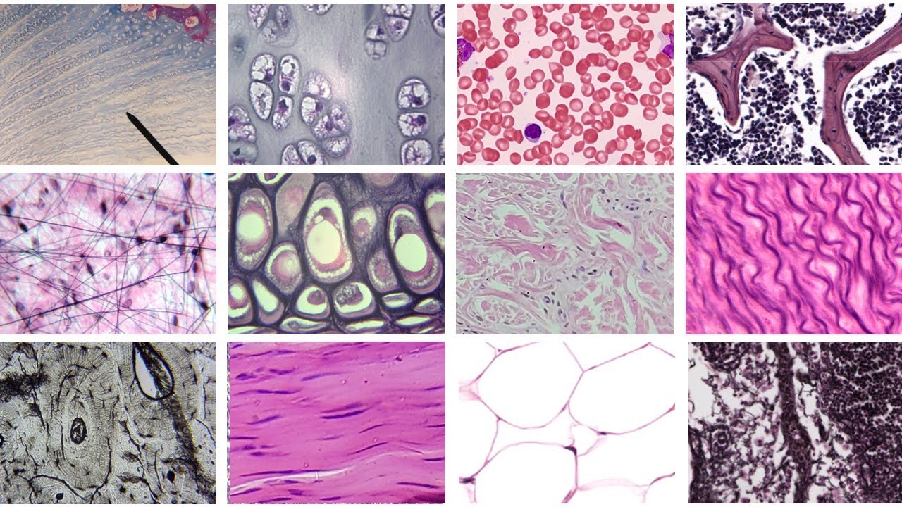
We already learned about cartilage when we discussed connective tissue, but let’s recall a few points. First, it contains a lot of water, which gives it the ability to withstand tension and compression. Cartilage contains cells called chondrocytes which sit in cavities called lacunae, inside of an extracellular matrix filled with ground substance and fibers.
There are three types, hyaline, elastic, and fibrocartilage, and skeletal cartilage is comprised of all three. Hyaline cartilage makes up the most skeletal cartilage, found in the nose, the ribs, the larynx, and the ends of most bones, providing support. Elastic cartilage is more stretchy, found in the ears, and the epiglottis, the flap that covers the opening of the larynx when we swallow.
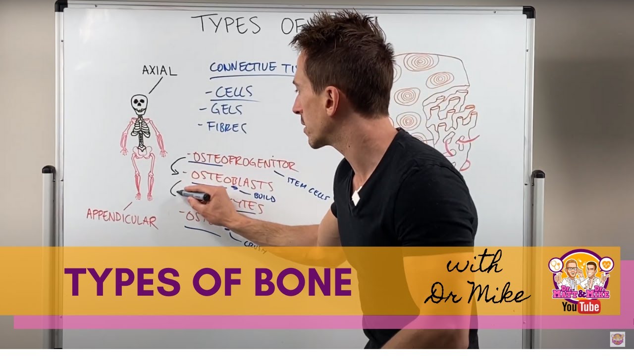
Then, highly compressible fibrocartilage can be found in areas that must withstand lots of pressure, like between the vertebrae of the spine. Now that we have the cartilage out of the way, let’s take a look at bones. Bones can be placed into two categories, axial and appendicular.
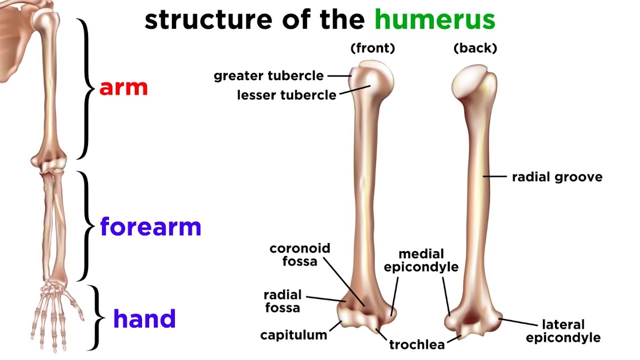
Axial bones are found in the head and torso, making up the spine, rib cage, and skull. Appendicular bones make up our appendages, or limbs, those being the arms and legs, as well as the pelvis and shoulders. Bones can also be classified by shape, being either long, short, flat, or irregular.
Long bones are longer than they are wide, like the ones in our limbs. Short bones are cubelike, found in the ankles and wrists among other places. Flat bones are thin and often curved, like the sternum and shoulderblades.
And irregular bones are the ones that have complicated shapes that don’t fit into the other three categories, like vertebrae and hip bones. What do bones do? Clearly the main function is support.
The rest of the body essentially hangs on the skeleton, supported by the bones as we stand and walk around. Also, organs are protected by bones, like the heart within the rib cage. Bones act as levers that allow us to perform physical tasks.
Bones also provide mineral storage, like calcium and phosphate, which can be released into the bloodstream as necessary, as well as fat storage, hormone production, and blood cell formation. So what exactly are bones made of? How can we connect this image with what we know about molecules and cells?
First let’s make the distinction between bone, a type of tissue, and an actual complete bone, which is an organ, because it is made of several types of tissue. Most of a bone is made of bone, but there is also nervous tissue, connective tissue, cartilage, and blood vessels. We will have to examine bones at a few levels of complexity, starting with gross anatomy, meaning the part that is visible to the naked eye.
The outer layer of any bone is made of compact bone, which is very dense and smooth. Inside there is lots of spongy bone, which is like a honeycomb of little needles. Typically the open spaces will be filled with bone marrow, which we will discuss in a moment.
The precise arrangement of compact and spongy bone depends on the bone type. Short, irregular, and flat bones all consist of thin plates of spongy bone covered by compact bone. There is no well-defined cavity for the marrow to sit in, and hyaline cartilage covers portions of the surface that are involved with joints.
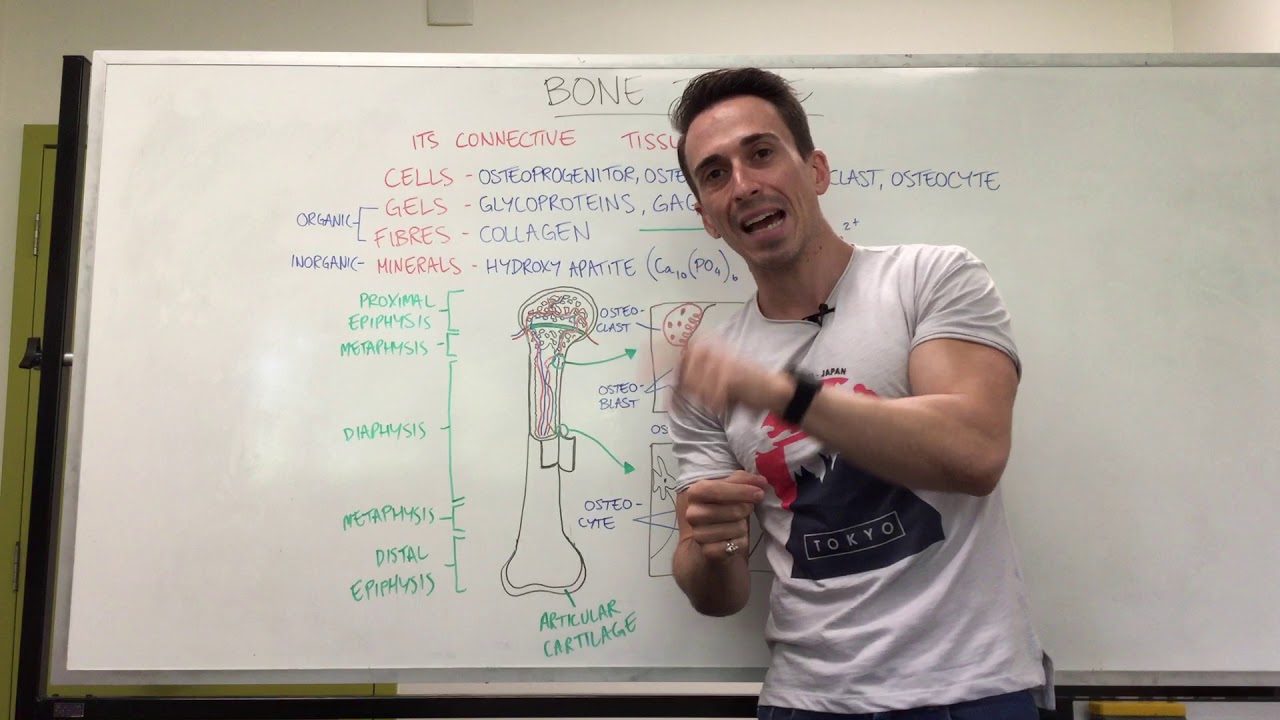
Long bones are a little different. These contain a tubular shaft, called a diaphysis. This is made of a thick collar of compact bone surrounding a medullary cavity, or marrow cavity.
In adults, this cavity contains yellow bone marrow which is high in fat. The ends of a long bone are called epiphyses. These parts do contain spongy bone inside the compact bone, and again, cartilage covers the joint surface for cushion and stress absorption.
Beyond the yellow marrow we mentioned, there is also red marrow, which can be found inside the cavities of spongy bone, and this type of marrow produces blood cells. We can also see an epiphyseal line, which is a remnant of the epiphyseal plate, a disc of cartilage that grows during childhood, which is how these bones get longer as a child gets taller. A white membrane called the periosteum covers the exterior of the bone, consisting of an outer fibrous layer made of dense irregular connective tissue, and an inner osteogenic layer, containing primitive stem cells.
This membrane is attached to a network of nerve fibers and blood vessels, which then pass through the shaft to the marrow cavity, and perforating fibers connect the periosteum to the bone. Endosteum covers the internal spongy bone layer, as well as canals that pass through the compact bone. In addition, the outside of a bone will display markings, which can be projections that bulge out, or depressions and openings like fossae, foramina, and grooves.
Now that we have this view covered, let’s zoom in a little more and check out the microscopic anatomy of a bone. We can find a few different types of cells in here, so let’s go through each one. First, osteogenic cells.
These are a type of stem cell that actively divide, and they are found in the periosteum and endosteum that we mentioned. If the bone is growing, these are flattened or squamous cells, and they can differentiate into other types at certain times. Next are osteoblasts.
These are the ones that secrete the bone matrix that consists of collagen and other proteins, meaning they are responsible for bone growth. These are also actively mitotic, and cube-shaped while active. Once surrounded by matrix, they become our next type of cell, osteocytes.
These are mature bone cells that monitor and maintain the bone matrix, communicating this information to other cells. Next are bone lining cells, which are flat cells found on the surface of the bone. These also help maintain the matrix.
And lastly, osteoclasts. These are large cells with multiple nuclei that use enzymes to break down bone, which is a normal process called resorption that releases minerals to be transferred to the blood. Now that we are familiar with the five types of cells, let’s zoom in on a long bone and see what’s what.
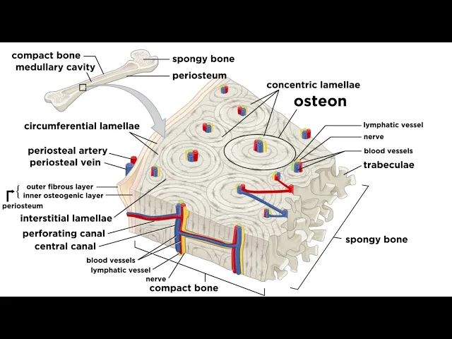
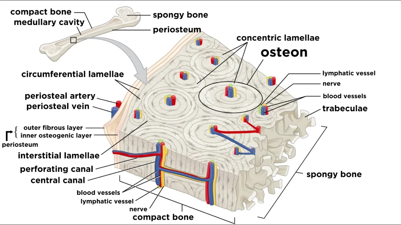
First let’s check out the compact bone in the shaft. This is actually not solid all the way through, there are many cylindrical units with open canals at the center, and each of these units is called an osteon. If we were to pull one of these out of the bone, we would see that it is made of a series of lamellae, which are hollow tubes, and these are arranged like the rings of a tree trunk.
Within each lamella are collagen fibers running in a specific direction, with crystals of bone salts in between, and as we proceed inward, the next lamella will have its fibers running in another direction, continuing in this fashion all the way to the center. This alternating pattern is what gives compact bone the ability to withstand torsion, or twisting force. The open region at the center is called the central canal, and it contains blood vessels and nerve fibers that serve the cells in that osteon.
There are also shorter canals running perpendicular, allowing for connections to run all the way from the periosteum to the central canals to the medullary cavity. Where the lamellae meet we can find tiny gaps called lacunae, and these are filled with osteocytes. The lacunae are connected by extra-tiny canals called canaliculi.
Beyond the lamellae found within osteons, there are others called interstitial lamellae, that fill in gaps between osteons, as well as circumferential lamellae, which make up the circumference of the diaphysis, surrounding all the osteons. Lastly, we can discuss the chemical composition of bone. There are organic components, which are all the cells we discussed, as well as osteoid, which is the organic part of the matrix.
This is made of ground substance and collagen fibers, which are secreted by osteoblasts. These materials contribute to the structure, flexibility, and tensile strength of the bone. There are also inorganic components inside any bone, such as hydroxyapatites.
These are needle-like crystals of calcium phosphate surrounding the collagen fibers, which largely accounts for the hardness of bone. The organic and inorganic components work together to ensure that bones are durable and strong without being brittle, and bones are almost as good as steel at resisting tension and compression, which is pretty miraculous when you think about it. Now that we know more about the structure of bones, let’s zoom back out and take a look at how they come together to produce an entire human skeleton.