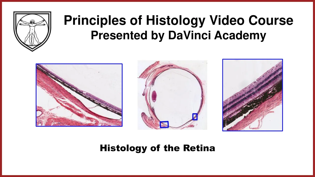
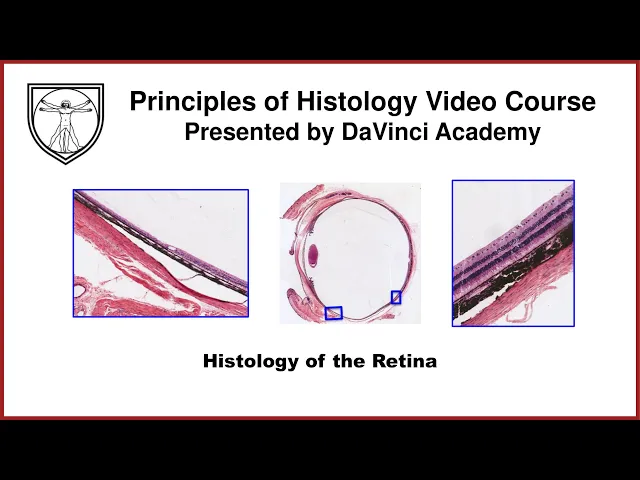
welcome to the da vinci academy histology video course the entire video course is available on youtube and covers all of the fundamental principles of histology and relevant cell biology you can find all the videos from the course by clicking the histology playlist link in the description below and then you can access the corresponding practice questions and histology lab videos by going to our website which is also linked in the description below welcome back in this third special census lecture we'll wrap up the histology of the eye by looking at the detailed histology of the neural
tunic of the eye in discussing the neural tunic histology it's actually helpful to look back on how this tunic develops embryologically because it is comprised of the two embryonic tissue layers which grew directly out of the developing brain so if we were to look at the developing brain in this crude diagram we'll see that there are these direct out pocketings of the brain on either side right and left side this is the anterior view where the bulbous end are called the optic vesicle and this narrowed region is called the optic stalk the optic stock will
eventually become the optic nerve and an interesting thing happens with the optic vesicle where the distal end starts to kind of cave inward like this once this optic vesicle takes on this cup-like shape surprise surprise we call the optic vesicle the optic cup now and at this point we can see that there are two tissue layers comprising the optic cup the outer and inner optic cup layers that are now separated by a little bit of this fluid-filled cavity called the intra-retinal space and it's the outer and the inner optic cup layers that are giving rise
to the neural tunic and as the inner optic cup layer grows thicker the intraretinal space will eventually disappear so the outer layer of this optic cup becomes the pigmented epithelium as cells in this layer start to accumulate pigments within its cytoplasm and the inner optic cup layer will eventually give rise to the sensory and non-sensory epithelium of the neural tunic and as these two layers mature and especially as the inner optic cup layer starts to thicken the intra-retinal space will eventually disappear in adults but interestingly enough this intra-retinal space is always going to be a
potential space because there aren't any strong cell cell junctions that are binding these two layers together so with a blunt force trauma to the head or to the eye these two layers can separate and that potential intraretinal space can reappear which is clinically known as the detached retina the neural tunic has three distinct histological regions parasitica is the neural tunic that's lining the interior of the iris so we've already seen this as we looked at the higher magnification of the iris so this is a stratified cuboidal layer of epithelium that is lining the inner aspect
of the iris we have also seen the next region of the neural tunic that is the parsiliaries this is the stratified cuboidal epithelium that line the inner aspect of the ciliary body and especially the ciliary processes to work along with the vascular tunic to produce aqueous humor the third region of the neural tunic is the most well known of the three that is the retina so this is the rest of the neural tunic that is responsible for the actual visual perception that is transported through the optic nerve to the brain and even from the macroscopic
view we can appreciate that the retina is quite thick towards the back but as we move towards the front or anterior aspect of the globe that retina layer starts to become thinner thinner thinner still and right around here is where the transition between the retina and parsiliaris may occur and this boundary between the retina and parceliaris is anatomically called the aura serata and this is because when we look at the globe from the posterior aspect from the inner aspect what we see in three dimension is almost a serrated outline between the retina and parsiliaris that
go all the way around hence the name aura surata and this anatomical landmark occurs due to the histological thinning if you will of that retinal layer from multiple cell layers to just a two layered stratified cuboidal epithelium let's review the histology of parse iridica again we've seen this before of the higher magnification of the iris we saw the vascular tunic component of the iris which forms the anterior component but the inner component or interior aspect of the iris we have this stratified cuboidal epithelium that is the extension of the neural tunic and forming the pars
iridica because the cells of this epithelium are heavily pigmented we can't really discern this clearly but in reality this is comprised of the two cuboidal cell layers and each layer is derived from the embryonic outer and inner optic cup layers that are heavily pigmented for the purposes of preventing any light from entering the eye through the iris so blocking out any light coming in through the iris so that the only light entering the globe is through the pupil we've also seen the histology of parsiliaris before when we looked at the higher magnification of the ciliary
body with the vascular tunic comprising the bulk of the ciliary body but the inner aspect is lined by the extensions of the neural tunic which we now know as the parsiliaris so to summarize it is the inner aspect of the ciliary body it is comprised of the two cuboidal cell layers each derived again from the embryonic outer and inner cup layers and it's really the outer layer that is pigmented and the inner layer unlike the taurus iridica is non-pigmented functionally speaking the two layers form the epithelial lining of the ciliary body and in particular these
ciliary processes and they participate in production of the aqueous humor now onto the histology of the retina this is the largest portion of the neural tunic and it extends from the aura serrata anteriorly and it extends posteriorly from there and as it extends posteriorly it becomes thicker and thicker aura serrata is the irregular boundary between the retina and the parse ciliaris right around there looking at the higher magnification of this transitional zone right around that boxed area at a higher magnification we can see the sclera on the outside and the vascular tunic or the uvl
or the choroid in the middle and this is an artifactual tear so we won't worry about that in real life the sclera and the uv would be close together at any rate interior to the choroid we have this two layered appearance of the neural tunic of the parsiliaris and right around here almost abruptly we have thickening of this neural tunic which is the beginning of the retina and it'll continue posteriorly and become thicker and thicker so when we look at the posterior aspect of the retina in this boxed area we can really appreciate once again
the sclera and the choroid abutting each other and again this is an artifactual tear and internally the retina starts from this dark single layered cuboidal looking cells inward like this so all of this is the retina that has thickened quite significantly compared to the anterior aspect of that retina regardless of which region of the retina we're looking at anterior or posterior there's some commonalities in the histology in that the outer layer of the retina is always going to be comprised of that single layer of pigmented cuboidal cells as we see here and then the inner
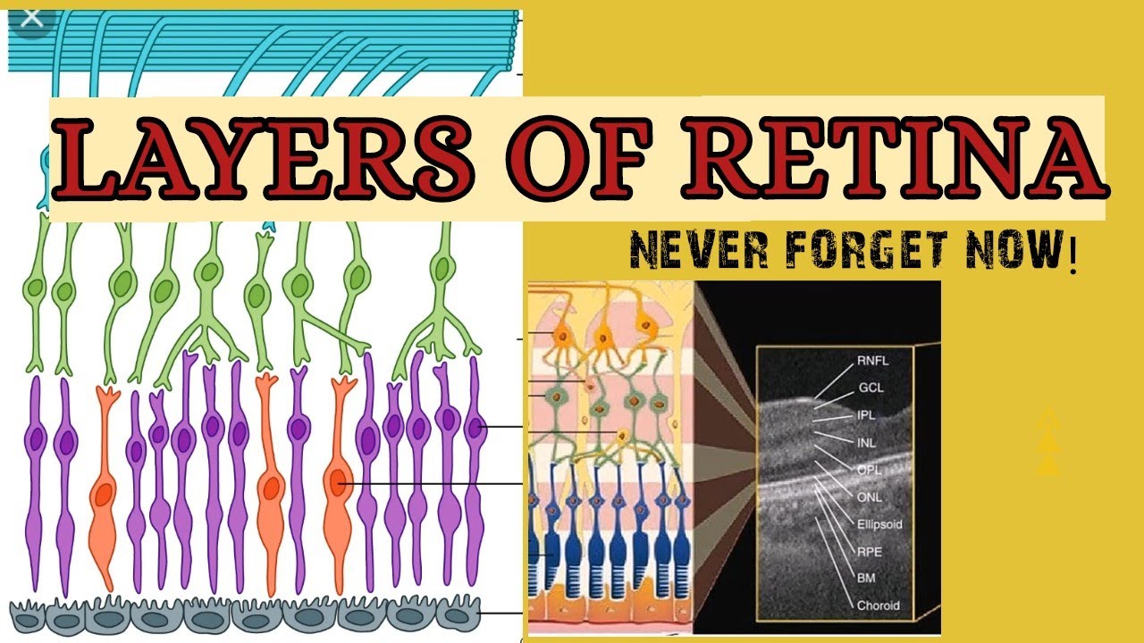
aspect or the inner layer which derived from that inner optic cup layer is much more thickened it's not pigmented and it is comprised of all the receptors neurons and glial cells that are reminiscent or consistent with the neural tissue the cells of the inner layer the receptors neurons and glial cells as well as the pigmented epithelial cells of the outer layer comprise the 10 distinct histological layers which becomes a lot more apparent as the retina itself becomes thicker towards the back to diagram this the external most layer is that outer pigmented epithelial cell layer that's
sitting on top of the brook's membrane which separates the retina from the choroid underneath the cells of the pigmented epithelium have a lot of these tight junctions towards the inner aspect these tight junctions are responsible for forming that blood retina barrier so anything that needs to be transported from the blood vessels of the choroid into the interior of the retinal tissue those things need to go through the pigmented epithelial cells rather than being able to go in between the cells another feature of these outer pigmented epidermium is that they have a lot of these microvilli
on their inner aspect and internally we have two different types of receptor cells these are specialized neurons with the receptor portions that are either looking like the rods or cones like these the receptors of the rods and cones are embedded within these microvilli of the outer pigmented epithelium but there are no cell cell junctions occurring between the receptors and the outer pigmented epithelial cells which means this is the intra-retinal space that used to be during the developmental process so this is where that potential space is and with any kind of mechanical blow or something like
that the entirety of the inner layer of the retina can lift off from that outer pigmented epithelium resulting in the detached retina the receptors of the rods are specialized to pick up the information on the light or the brightness of the environment as opposed to the cones there are three different types of cones in our eyes for most people and these are activated by specific wavelength of the light that's coming in so these are responsible for our color vision the inner aspects of these rods and cones have their exons branching out interiorly deeper in we
have specialized neurons called bipolar neurons that have their dendrites that come out from one pole and their exon branching out from the other pole so you can see that the dendrites of these bipolar neurons synapse with the axons of the rods and cones so should there be any action potential triggered they can then be relayed through these bipolar neurons to their exons more internally we also have third type of neurons that are even more interiorly positioned and these neurons are called the ganglion cells they too have their dendrites that synapse with the bipolar neurons and
they send out their exons that tend to go out and become quite long these axons will coalesce and exit the eye at the optic disk and collectively form the optic nerve which travels all the way to the brain there are a number of glial cells that support these different types of neurons of which we'll mention just one and that is the mueller cells these mular cells are fairly tall in fact they spend the entire thickness as you can see here of the entire inner retinal layer they surround all of these neurons and their extensions therefore
these mueller cells provide a not only the structural support but also they regulate and support the microenvironment of all the neurons in the retina muller cells nuclei are typically positioned around at the same level as the nuclei or the cell bodies of the bipolar neurons another notable thing about these mular cells are that they stop right at the neck of these receptor regions of the rosin cones so the mueller cells make cell cell junctions with each other as well as with the terminal ends of the rods and cones receptors and in this way mueller cells
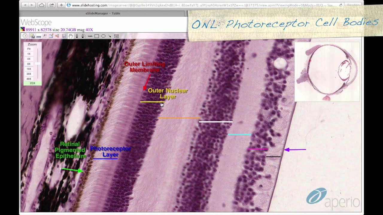
also contribute to the blood retinal barrier as well so let's see how these cells are actually making up 10 different histological layers going from external to internal here's number one the outer pigmented epithelium the second layer is right here which is comprised of the receptor regions of the rods and cones so that's number two number three is a really really thin boundary right here that is made by the cell cell junctions of the mueller cells with each other as well as with the base of the rosin cone receptor portions so this layer is called the
outer limiting layer or external limiting layer number four is right here and here on the diagram and this layer contains the cell bodies including the nuclei of the rods and cones so a lot of these heterochromatic dark circles are the nuclei that belong to the rods and cones so this is the outer nuclear layer comprised of the cell bodies of the rods and cones the next layer n is right here and in our diagram it's located right here and this a nucleate layer is where the synapse between the axons of the rods and cones and
the dendrites of the bipolar neurons are occurring so that's the outer plexiform layer is number five number six is located right here and on this diagram right here so this is another nucleated layer a lot of these nuclei are belonging to the cell bodies of the bipolar neurons as well as the nuclei of the mueller cells are positioned in this layer which is known as the inner nuclear layer moving even more inward we have number seven right there which corresponds to this layer in the diagram number seven another a nucleate layer and this is where
the synapse between the axons of the bipolar cells and the dendrites of the ganglion cells are occurring so this layer is known as the inner plexiform layer number eight right there corresponding to this layer in the diagram so this is the nucleated layer again and these nuclei belong to these ganglion cells so this layer is known as the ganglion cell layer number nine right here another a nucleate layer corresponding to this layer in the diagram that's number nine and as you can see from the diagram this is a layer of the axons of the ganglion
cells that are all coalescing and traveling together so this number nine layer is known as the exon fiber layer coming from the ganglion cells and finally here's number 10 indicated by the innermost aspect of the mueller cells and the name of this layer is the inner limiting membrane so these are the 10 layers of the retina let's summarize going from external to internal we have the pigmented epithelium right here then we have the receptors of the rods and cones layer here number two outer limiting membrane this really thin line between the receptors and where the
cell nuclei lie so this is the outer limiting membrane where the muller cells are making cell cell junctions with each other as well as with the rods and cones next layer in we have the outer nuclear layer and these nuclei belong to the rods and cones next layer in is the a nuclear layer of the outer plexiform layer the synapse between the rosin cones and the bipolar cells the inner nuclear layer makes up the sixth layer of the retina and these nuclei belong to the bipolar neurons as well as those of the glial cells such
as the mueller cells next layer in another a nucleate layer of synapse between the bipolar cells and the ganglion cells so this is the inner plaxiform layer right here next layer another nucleated layer of ganglion cell layer followed by the exon fiber layer all the exons emerging from the ganglion are traveling within this layer and lastly we have the inner limiting membrane which is the innermost boundary of the mueller cells that are once again forming cell cell junctions with each other so imagine i'm a molecule of photon coming into the eye i would actually have
to travel all the way through these layers in order to excite the receptors of the rods and cones then the action potential will travel back up towards the inner aspect of the retina and travel through the exon fibers and emerge out of the eye at the optic disk now while the 10 histological layers of the retina remains more or less consistent throughout the retina there are some specialized regions within the retina that's worth noting for example we have been seeing this dividend from the beginning this little dibben is called the phobia centralis and as you
can see from the macroscopic view this phobia centralis is in direct line of that light coming in through the cornea the pupil and through the lens so this is the area of the greatest acuity so whatever of the world we want to see at the highest resolution we would focus that image right onto the phobia centralis here the 10 layers of the retina are attenuated to allow for the photon to excite the receptors without too many interference this is also the area of the highest concentration of cones there are essentially no rods in this area
to allow for the greatest acuity of color vision if you will let's focus on this area and see its histology at higher magnification let's start with the orientation here's the vitreous humor here's the retina here's the choroid and here's the sclera we still see the pigmented epithelial layer and surrounding the phobia centralis we see this beautiful 10-layered organization of the retina however at the phobia centralis we see the layer comprised of the cone receptors the outer limiting membrane as well as the outer nuclear layer hosting the cone cells nuclei but the inner layers of this
area are greatly attenuated to the point where we can't clearly discern the inner nuclear layer the plexiform layers and ganglion cell layers so the photon doesn't have quite as many layers to get through to excite the receptors of the cones some clinical pearls associated with the retina and the phobia centralis we have the myopia which is the nearsightedness this is where our eyeballs are a little too long or elongated so the image that we're trying to focus on actually gets focused in front of the phobia centralis on the other hand in hyperopia or farsightedness the
eyeball happens to be a little too short so whatever we're trying to focus on gets focused behind the phobia centralis so whatever we're trying to focus on appears a little bit fuzzy so this is where we use either convex or concave lens to correct for these deficiencies and lastly the specialized area of the retina is the optic disc which is shown right here to orient ourselves here's the sclera choroid and the 10 layered organization of the retina on either side of the structure here we can see that disruption of the 10 layered retina but instead
what we see are all these axons from the axon fiber layer coming from the ganglion cells coalescing onto this single point called the optic disc and all of those axons exiting the eye and comprising the optic nerve so this is the site of all the ganglion cell exon fibers merging into one point in order to exit the eye and as you can imagine this is the area that's lacking any photoreceptors so this area is also known as the blind spot thank you for watching this video from the davinci academy histology video course which is completely
available on youtube to access the corresponding practice questions and histology lab videos go to our website using the link in the description below foreign foreign [Music] you
![Ear Histology [Special Senses Histology Part 4 of 4]](https://img.youtube.com/vi/hTeIRe2cBDQ/maxresdefault.jpg)
![Eye Anatomy and Histology of the Outer Layers of the Eye [Special Senses Histology Part 1 of 4]](https://img.youtube.com/vi/u1fUUr1A34Y/maxresdefault.jpg)

![Histology of Eye: Lens, and Mechanism of Accommodation [Special Senses Histology Part 2 of 4]](https://img.youtube.com/vi/_L255O4oefs/maxresdefault.jpg)
![Skin Histology [Integumentary System Histology Part 1 of 2]](https://img.youtube.com/vi/NBEjKRuGHjY/maxresdefault.jpg)
![Histology of the Ovary and Ovarian Follicles [Female Reproductive Histology Part 1 of 2]](https://img.youtube.com/vi/cDs6goPOD1I/maxresdefault.jpg)


![Pituitary Gland Histology [Endocrine Histology 1 of 2]](https://img.youtube.com/vi/XZrS4zkrSps/maxresdefault.jpg)