hello and welcome to another invigorating narrated lecture this time it's going to be over chapter 21 over adaptive immunity I will be breaking this up into at least two narrated lectures so that the files don't get too big all right let's go ahead and get started the first thing I want to do is actually introduce HIV um as a virus and this the reason why I want to do this is twofold first of all I want you to see how HIV is um capable of completely crippling the immune system and second I want you to
see um the role of te- cells as so this is an example see the role of T cells all right so the structure of HIV is shown in panel a it's an enveloped RNA virus and it has a variety of proteins on its surface the one that we're interested in is gp1 so it's a spike protein called gp120 and gp120 is able to bind and recognize a receptor protein receptor on the t- cell that's what you see in panel B once this happens the membranes will fuse and the nucleocapsid enters the T cell and it
has been successfully infected now to see why this is such a bad thing um let's look quickly at how te- cells function just very very briefly in general um here at the top we have a dendritic cell dendritic cells as you know from the previous chapter they are part of the inate immune system they are fosic cells that also are very good at presenting antigen so they are very important antigen presenting cells and that's exactly what they're doing here uh this this dendritic cell is presenting antigen to two t- cells one is shown in green
and is labeled as cd8 one is shown in blue and is labeled as CD4 you will understand these designations later but for now we're not so worried about it um now once these cells are activated you can see that they go off and activate many other cells in the immune system so cells of the innate immune system and other cells of the Adaptive immune system like V cells which make antibodies and um other cells that end up killing infected or tumor cells so you can see here if HIV infects and ends up decreasing the number
of te cells in the body we completely the entire immune system not just the Adaptive Branch but both branches because t- cells are important coordinators between both speaking of adaptive versus innate immune systems um I want to just go through a couple qualities of the Adaptive branch and compare them to the inate immune system in the process so we have a lot going on in this slide um first of all I want to point out that the Adaptive branch is very specific to a pathogen um that is it's able to and needs to recognize very
specific epitopes on antigens so let's back up a minute and Define these two words so an antigen as we've discussed before is a little molecular tag and an epitope is just simply a small region of that antigen very specific three-dimensional region on that antigen um proteins Serve by far as the best antigens because they are so variable um there's so many Poss possible structures that they can make so many potential threedimensional confirmations let's compare that to carbohydrates and lipids those don't serve as very good antigens they are very redundant there's a lot of repetition in
their structure and um so the innate immune I'm sorry the Adaptive branch of the immune system when it recognizes antigens uh tends to stay away from Pure carbohydrates or lipids or if they do bind these um maybe not as we don't get as strong of an an immune response okay so the take home message here um from from this bullet point is that proteins make the best antigens and often the proteins are bound to carbohydrates and lipids which again enhances their ability to be recognized by the immune system okay so again the Adaptive branch is
very specific to these epitopes let's compare that to the innate Branch um remember that the innate branch of the immune system is very non-specific so it doesn't care what micro we're talking about it serves as a Frontline barrier physically and chemically and with cells um they're they're not discriminatory okay um okay back to the Adaptive brand here uh how does it discriminate between these antigens well the how is actually kind of a lengthy answer that's still up for debate um but one thing I want to point out with this concept of the ability of the
Adaptive Branch to discriminate bad versus good uh is that there's a kind of a widely accepted Theory out there that the immune system is able to to see something that is self versus something that is nonself so something is part of you versus something that's not a part of you so the self is considered good and okay and the non-self is considered bad and not okay potentially um there's a lot of debate about this though and if you remember from the last chapter Dr matzinger came up with this idea called the danger Theory the danger
hypothesis um which states that indeed cells are actually looking for danger signals rather than self versus nonself antigens so we'll kind of need to keep this in mind even though throughout this chapter I will be referring to self versus nonself occasionally um finally as far as the discriminatory quality of the Adaptive demun system I want to bring up this concept called immunogenicity immunogenicity is the ability of an antigen to generate an immune response as I mentioned before proteins do this the best but something called a hapton is actually too small to generate an immune response
so it is not immunogenic at all um however these small little molecules again referred to as hapton are often conjugated or can be conjugated to a carrier molecule maybe it's a plasma protein that it binds to and now it is immunogenic so good example of this would be penicillin the drug penicillin an antibiotic that some people are highly allergic to there um therefore that means that their immune systems recognize recognize as bad and launch an immune attack um this is because because penicillin is even though it's a hapon it binds to plasma proteins and therefore
is suddenly immunogenic that's what's going on in these people okay and finally the last thing I want to mention as far as um these qualities of adaptive immunity is the idea that it has a memory so if you become infected with a particular pathogen you will generate um some memory cells some memory B cells and T cells that can act very quickly should you become infected later down the road with the exact same pathogen so the um second time that you're infected with the same pathogen these memory cells can launch a stronger immune attack much
faster before the pathogen has chance to take hold and you might not even realize that you were ever infected in the first place let's look at this graphically okay so um even though we haven't talked about antibody structure or anything yet about antibodies uh let's just take the uh the names here for granted so you see several traces on this graph okay uh you see IGM and IGG in both cases so on the left here this is our primary antibody response meaning this is the first time you were infected with this particular pathogen uh you
see that we have a certain type of antibody in the class called IGM IGM antibodies are very uh produced uh first before IGG uh and you know even despite their production it takes about a week before we see this um compare that to the secondary response so the second time you become infected with this same pathogen it only takes maybe one or two days before we we have antibody production and also notice that IGG just takes off so we have a stronger response both IGG and IGM are very effective at activating our immune system and
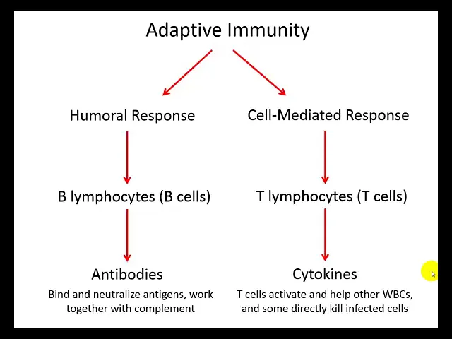
clearing these microbes from the blood now while we're talking about this idea of memory let's one more time go back to our innate immune system and remember that it has no memory okay um it is a Frontline defense that's not specific and it has no memory component so you could be reinfected with the same thing it doesn't matter because it doesn't discriminate at all okay so here's sort of the outline of this chapter um adaptive immunity can be subdivided into two major branches let's take the left Branch first the so-called humoral response is the part
of the adaptive branch that involves antibodies In The Blood and Tissue fluids so humor refers to body fluids um most of the time we be talking about the blood but this could also uh include other body fluids like tissue fluid uh this Branch primarily involves B cells B lympocytes and once activated they will produce antibodies um the other branch of the immune response is the cell mediated Branch this is where te- cells are primarily involved and rather than secreting antibodies they secrete cyto kindes to coordinate all the cells of the immune system um they're going
to activate other lucites and some of these lucites will directly kill infected cells okay so uh hence the sort of the name here cell mediated response this is where we have cell to cell contact the at the end result here cellto cell contact where those cells are ended up those infected cells are end getting killed okay U one last thing I want to mention here is that these branches are not entirely independent of each other the T cells for example are going to be involved with activation of the B cells uh through these cyto kindes
okay so the first thing we're going to do is we're going to take the humoral Branch we're going to talk talk about B cells and antibodies first before we get into the dell the details of B cells and their activation I want to first talk about antibodies what they are what they do what they look like and how they're made um an antibody is the same thing as an IM immunoglobulin so you will hear it either way these are y-shaped protein molecules consisting of four polypeptide chains you can see this down here on the bottom
left we have two heavy chains in the middle they're the long ones and then we have two light chains those are the short ones all these chains are connected to each other by dulfi Bridges so these sulfurs that are part of the side groups of the amino acids in these polypeptides form these so these disulfide bonds okay so it's just kind of kind of f everything together all right now notice that the tips of the y-shaped molecule are different color and the reason why they are different color is because these are the regions That Vary
a lot in their structure so they're the variable region or the variable domain of the antibody molecule and these are also the regions where antigens are bound okay so each antibody each y-shaped molecule here can B up to two antigens or epitopes keep in mind that these variable regions on a single antibody are going to be identical so this antibody is going to bind one specific epitope it's not going to bind two different epitopes all right because there is a huge diversity of epitopes out there in existence we have to have a huge variety of
variable region confirmation as well so in a little bit we're going to be talking about how these antibodies are formed through this mixing and matching process with their genes in order to get a huge variety of shapes uh and confirmations so that they can hopefully bind all those antigens in uh that exist out there okay let's move down now to the constant region or I'm sorry the FC stem here the FC stem of the y-shaped antibody uh is composed of the constant domain of heavy chains and the FC stem as we'll see a little bit
later is going to be very exposed when the variable regions are bound to a pathogen so if the variable regions are bound to a pathogen the FC stem is exposed to fos sites it's also exposed to uh complement proteins and can activate both over here on the right we see rather than kind of this block cartoon um on the left on the right we actually see a three-dimensional molecular model and you see that it still sort of retains that y-shaped confirmation all right in this picture here I want to try to tie in some stuff
some stuff that you've potentially learned in BIO 202 with regard to the utenation reactions in the blood type system okay so in panel a there's a red blood cell with a particular antigen called antigen a so this person that whoever contains this red blood cell uh this person has Type A blood and notice that um in this picture we have two types of antibodies an antib and anti-a if you look at antib antibody for just a moment you see a bunch of y-shaped monomers individual y-shaped antibodies that are stuck together in a penter so we
have five of them stuck together and some one class of antibody in particular often comes in this way so we have up to 10 binding sites then in this pentamer all right so whether it be anti-a or anti-b we have that same kind of penter shape with the variable regions poking out so if this is antigen a on the arthrite surface then anti-a antibody is going to be very specific for that antigen and if it should bind then we have the result in panel B because we have multiple binding sites on these anti-a antibodies uh
the antibodies can bind and sort of link a bunch of erthrocytes together causing this clumping or in other words utenation if we look at this under the microscope you see normal in panel C and uated in panel D now you can imagine rather than red blood cells you can imagine this being a pathogen of some sort SW and this is one way that antibodies work is that they cause stuff to Clump if these were a bunch of bacterial cells and we cause them to Clump together a couple things are going to happen first of all
that's going to reduce the ability of the microb to move throughout the body and infect um other tissues and second um it's going to serve as this giant Target for phagocytosis so this is one example of how the structure of an antibody kind of complements its function as far as clearing pathogens from the body okay now we have our classes of an antibodies here there are five of them and I want you to be familiar with each one as well as some of the properties of each so let's start with IGG and IGM because they
are um probably they're they're uh the two types that we see during a primary antibody response and a secondary antibody response as we as as we saw in previous graph all right IGG first um IG is by far the most abundant in the blood um it lives the longest and it's pretty small functions as a monomer therefore it can cross the um endothelial cells uh it it can cross the uh blood vessel barrier okay so so these IGG monomers that are circulating through the blood can actually exit into the tissues now likewise they could do
the same thing passing from maternal blood to fetal blood uh if we're talking about crossing the placenta so they are able to do that and these are the only ones that can cross the placenta um IG does a good job of of neutralizing microbes and toxins by causing them to kind of clumping them together but also um preventing them from binding their host receptors so if we take a virus as an example if we bind the virus with IGG then the virus is not going to be able to um penetrate its host cell IGM is
the largest antibody of the five classes in fact its name implies that with M meaning macro so IGM is the largest because it's so large as a penter it's not able to cross um through the blood vessel wall okay so it remains in the blood IGM is also quite shortlived compared to IGG as we saw in the graph a little bit ago migm appears first in response to a primary infection but second in response to a secondary infection or or a subsequent infection with the same microb remember that IGG appeared second but in higher numbers
with a primary infection and with a subsequent infection with the same microb it appeared second and in much higher abundance than IG IG is involved with clumping things together sort of neutralizing our um our pathogen uh activating compliment it's one thing I forgot to mention with IGG it activates complement as well in fact those are the only two that activate compliment and IGM is also involved in the transfusion reactions with the blood types as I showed you on the previous slide up next is IG IGA is uh it can be found as a monomer or
a dimer you see the dier shown down here on the bottom left of the screen Diemer has two monomers stuck together with this protein this joining chain this Joiner in the middle here and then a secretory component which in this cartoon looks like this uh snaking yellow protein and that's going to help prevent this molecule from being destroyed so IGA is not very abundant in the blood when it is in the blood it's found as a um a monomer and its function is not well known but as a dimer it is secreted into um the
mucus membrane as well as other body secretions like tears so IGA kind of plays a preventative role um it's its main goal is to neutralize the microb and prevent it from actually attaching to the host and entering the host now even though it's not very abundant in the blood it's super abundant the most abundant imunoglobulin in uh the mucus membranes and it's so abundant in fact that if we take all immunoglobulins together it turns out that IG is actually the most abundant in the body um so be careful here because IGG is the most abundant
in the blood IG taking everything into account IG is the most abundant in the body just because there's so much produced and secreted in those mucous membranes okay next up is igd there's not too much to say about igd it's an immunal globulin that serves as a receptor on V cells so we will come back to that a little bit later um it's rolling the serum if any is not well known IG last but not least by far the the lowest abundant um it's far less than 1% of total immunoglobulins found in the blood uh
it also serves as a receptor on basophils and other cells involved with allergic reactions like mass cells and IG is also uh involved with fighting parasitic infections so this table summarizes everything that we just talked about here as far as what it looks like like what's called um its relative abundance its halflife simply refers to its lifetime in the blood um whether or not it can activate compliment notice that IGG and migm are the only two that can activate compliment the other other ones cannot uh IGG is the only one that can cross the placenta
and because IG and igd serve primarily as receptors they don't do a good job job of actually neutralizing our pathogens and toxins but the other three classes do okay all right now let's talk a little bit about how antibodies are produced I mentioned that the number of antigens possible out there um in the world is mindblowing talking about um you know every possible molecular tag there's there's a ton of possible um sequences and three-dimensional shapes for proteins for example so our immune system in order to recognize all these epitopes on antigens uh also has to
be good at producing a bunch of variety um it turns out that our immune system is actually pretty good at this though despite the odds so um it's estimated that our immune system has the ability to respond to the majority of the possible epitopes out there up to maybe 100 trillion or more now what's kind of crazy about this is that all of this diversity is produced by the mixing and matching of only about 600 Gene segments pieces of genes that are are sort of shuffled around during this process called sematic recombination um this is
also known as vdj Rec combination and you'll see why on the next slide so we get a lot of variety through this mixing and matching and putting them in different orders we also get some variety through mistakes that are made as well as mutations all of those are going to result in increased variation in our antibodies and other things okay um this somatic recombination is occurring primarily in the bone marrow for B cells and thymus gland for t- cells those cells are then later going to leave and enter lymphatic tissue Etc but while they're here
in the bone marrow and the thymus they develop um they develop their receptors through this bdj Rec combination and they are tested against body cells so this is where we come back to that self versus nonself idea we don't want B cells and t- cells that end up recognizing our own antigens as non-self or bad antigens we don't want them to launch an immune response against our own cells so in order to avoid that these B cells and T cells cells are screened in a process called clonal deletion if they should react to self antigens
they're killed immediately so through this clonal selection process a very small percentage actually make it through and then of that small percentage we're supposed to recognize all of the possible for epitopes that exist so we definitely have our work cut out for us here at the immune system now the last thing I want to point out here is that we're doing this mix mixing and matching and this genetic recombination uh for a few things V cells and t- cells both have receptors that will need to be re able to recognize um epitopes of pathogens therefore
they need to have all that variety so The receptors for B cells and T cells both undergo this sematic recombination process when they're being produced in addition we have antibodies that are secreted and released into the plasma all these different antibodies like IG and IGM they're released from activated B cells as we'll see later and um when these are made uh they're of course going to be um ultimately their structure stems back from this Ming mixing and matching uh somatic recombination okay again the details might be a little bit fuzzy right now but we're going
to we're going to go into all the nitty-gritty here shortly when we look at B SEL activation okay but for now I just want you to keep in mind that The receptors on B cells and T cells undergo this sematic recombination and then this screening process in the bone marrow and the thymus to determine um to select for clones that don't react for self okay here is what I was just saying in pictures so we have an IM imunoglobulin kind of in the center at the bottom that's made we have the heavy chains the long
polypeptides shown as Gene segments are shown on the left and the light chains Gene segments are shown on the right let's take the heavy heavy chain because the light chain is very similar so notice that we have several different Gene segments v d j and C so the v stands for variable this is going to be um definitely part of the variable domain and we have up to 300 Gene segments for the V Gene we have 30 different D genes and d stands for diversity we have up to six different J or Joiner genes and
we have eight different C or um constant domain genes so in order to make our heavy chain we need to pick one of each so we pick one of the V one of the D one of the J one of the C and it doesn't matter what combination just sort of this mixing and matching idea and we get it we're going to get a huge possible variety of variable domains as a result okay now that we understand how what antibodies are how they're made we also um want to understand what they do okay so how
antibodies act as eors how they go off and do something in the cell um earlier I showed you how uh antibodies can neutral neutralize a toxin or neutralize a pathogen by causing this utenation or clumping to occur and that's um that's what we have here shown in panel D so let's just start with where we've come from okay so panel D here utenation that's review for you in this case we see that rather than red blood cells though these are bacterial cells okay so clumping of the bacterial cells called glutation is going to prevent their
Mobility uh restrict their mobility and it's going to prevent attachment to their host cells or tissues we have the same kind of thing that can happen with small soluble dissolved antigens um these antibodies can sort of Crosslink those and this is going to serve as a target for phagocytosis all right let's go up to A and B A and B are basically the same thing so rather than worrying about viral inhibition we're just going to call both of them neutralization so neutralization the antibodies are binding the surface and notice that they're not directly killing the
the virus in this case and they're not directly destroying the toxin they're just simply binding to it and preventing its ability to enter our cell and cause disease okay so this is neutralization and then finally in panel c um when an antibody binds a microbial surface like this bacterial cell variable domains are stuck to the antigens on the surface of the cell but the FC stem is poking outward and I was referring to this a little bit ago the FC stem is recognized by fyes and um enhances phagocytosis so this in this way the antibodies
are serving as obstinance so we're slathering on the butter here enhancing the attractiveness of that bacterial cell by this fosite