Hello, I m Dr David Spach from the University of Washington. In this National HIV Curriculum Mini-Lecture, I'll provide an introduction to the biology of HIV-1. This talk will begin with an overview of the main structural features of HIV, followed by a brief look at the HIV genome.
Then I'll take a more in-depth look at each of the HIV proteins, including structural proteins, enzymes, and the accessory and regulatory proteins. The overview of these five topics will hopefully provide you with the foundation to understand more complex HIV-related topics, such as the HIV life cycle and the mechanism of action of antiretroviral medications. This talk will not address HIV-2, and in the following discussion, any mention of HIV will be in reference to HIV-1, which is the dominant HIV type found globally.
Let's start first with a brief look at the basic structure of HIV. HIV is a retrovirus that is roughly spherical in shape and only about 100 to 150 nanometers in diameter. To put this in perspective, a nanometer is only a billionth of a meter.
The outermost part of this virus is the envelope, which consists of the envelope glycoprotein spikes and a bilayer lipid membrane. Just inside the membrane, HIV has a large matrix shell, which is comprised entirely by the HIV matrix proteins. Moving inward, HIV has a protective core, which is commonly also called the viral capsid or capsid core, and this cone-shaped structure consists of clusters of the HIV capsid protein.
The innermost structural component of HIV is the nucleocapsid complex, a tightly woven mix of HIV RNA and several other proteins. In this next section, I'll briefly examine the HIV genome. Each virion contains two identical copies of single-stranded genomic HIV-1 RNA.
The genomic HIV-1 RNA is a positive-sense strand that is approximately 10,000 base pairs in length. The entire HIV-1 genome contains only nine genes; these nine genes encode for the downstream production of a total of 15 proteins. Next, I m going to focus on the HIV proteins.
To better understand the role of each of the 15 HIV proteins that are produced, it s useful to group them into four main categories: structural, enzyme, accessory, and regulatory. There are six structural proteins: gp120 and gp41, which together make up the HIV envelop protein, matrix, capsid, nucleocapsid, and p6. There are three enzymes: protease, reverse transcriptase, and integrase.
There are four accessory proteins: vif, vpr, vpu, and nef, and two regulatory proteins: tat and rev. These 15 proteins have distinct functional roles for HIV. And they vary significantly in size.
This image shows the relative size of the HIV proteins based on the number of amino acids in the mature protein. In this illustration, I grouped the gp120 and gp41 subunits together as a single HIV envelope glycoprotein, and the HIV protease is shown in its functional dimer conformation. Next, let's take a closer look at each of the HIV structural proteins.
The major HIV structural proteins are envelope, matrix, capsid, nucleocapsid, and p6. The outermost structural protein is the HIV envelope glycoprotein. Each HIV particle has about 10-15 of the envelope glycoprotein spikes that are randomly arranged like little knobs on the viral surface.
Each envelope protein includes the gp120 and gp41 subunits. The gp120 subunit is also referred to as the surface glycoprotein, and the gp41 is the transmembrane or membrane-spanning glycoprotein. The envelope protein has an overarching trimer structure with each trimer consisting of three gp120-gp41 pairs.
Each of these gp120 and gp41 pairs is joined by a noncovalent bond. The envelope glycoproteins play a critical role in HIV entry into the host cell. Two regions on the gp120 subunit--the CD4 binding region and the V3 region--are involved in the early steps of viral entry, whereas gp41 is involved in the later stages of viral entry.
The gp120 subunit is also a key target for the host immune responses against HIV. As shown here, the outer surface of gp120 is heavily glycosylated. This glycan shield effectively cloaks many of the critical immune determinants that are on the surface of gp120, which helps HIV to evade host immune responses.
Now moving inside the viral lipid membrane, let s examine the HIV matrix (or p17) protein. In each virion, there are about 2,500 matrix proteins. These proteins are attached to the inner surface of the HIV bilayer lipid membrane.
The matrix proteins have a wider N-terminal domain that is closest to the cell membrane and a more narrow distal C-terminal domain. The N-terminal domain contains a highly basic region--shown in purple here--that initiates binding of the matrix protein to the host cell membrane during viral assembly. The N-terminal domain also contains a myristyl group--shown in light blue--which can transition between two conformations.
At baseline, the myristyl group is in a sequestered conformation, but when the matrix protein gets close to the cell membrane, the myristyl group can spring out into an exposed conformation. The exposed conformation promotes binding and anchoring of the matrix protein with the membrane. In the mature HIV particle, the matrix proteins are grouped as trimers bound to the bilayer lipid membrane.
Just beneath the lipid membrane, these matrix trimers interlock to form an intricate spherical lattice-like structure that's referred to as the matrix shell. The matrix shell plays an important role early in the HIV life cycle by facilitating HIV entry into the host cell. Matrix also plays a role late in the HIV life cycle during viral assembly.
When matrix is synthesized, it is initially part of the HIV gag and gag-pol polyproteins. During viral assembly, the matrix protein anchors these large polyproteins to the inner region of the host membrane. Let's move further inside HIV and take a look at the HIV capsid (or p24) protein.
The HIV capsid protein makes up the outer structural element of the cone-shaped HIV core, which again is also referred to as the HIV capsid or HIV capsid core. Each core contains about 1,500 capsid proteins. The capsid protein has two major domains that are connected by a flexible linker.
This flexible linker plays an important role when these capsid monomers begin to bind together and assemble into larger clusters. As shown here, the capsid monomers assemble into polymers, eventually forming groups of six hexamers and groups of five pentamers. The formation of hexamers occurs about 20 times more frequent than the formation of pentamers.
The hexamers and pentamers further assemble to form the cone-shaped HIV core. This complex assembly process occurs very late in the HIV life cycle. The fully formed HIV core has a cone-shaped structure that contains about 250 hexamers and 12 pentamers.
The HIV core provides a structural barrier that sequesters and protects the HIV RNA, and key enzymes, and several of the accessory proteins. During viral replication inside the host cell, the primary role of the HIV core is to deliver the contents within the core from the cytoplasm to inside the host cell nucleus. Now let's move even further inside the HIV particle to look at the nucleocapsid (or p7) protein and the ribonucleoprotein complex.
The HIV nucleocapsid protein coats the HIV RNA. Each strand of genomic HIV RNA is coated with about 900 HIV nucleocapsid proteins; each nucleocapsid protein covers about 11 to 12 base pairs of the genomic HIV RNA. HIV nucleocapsid is a small basic protein that contains N-terminal and C-terminal regions and two zinc-binding domains that recognize and bind to specific sites on the HIV RNA.
Most of the nucleocapsid proteins are tightly complexed with the genomic HIV RNA. The complex of HIV RNA and nucleocapsid protein is often referred to as the ribonucleoprotein complex. Some more recent models also include the HIV integrase enzyme as part of this ribonucleoprotein complex, with the integrase proteins predominantly grouped as dimers or tetramers.
These images show a more realistic rendition of the HIV ribonucleoprotein complex, as shown without and with the HIV integrase enzymes. The nucleocapsid protein plays an important role in multiple steps of HIV reverse transcription. The last structural protein to discuss is HIV protein 6 (or p6).
HIV p6 is a very small protein, and the role of free p6 and the mature virion remains unclear. This protein has N-terminal and C-terminal regions connected by a flexible region. This flexible region enables p6 to serve as a docking site for other proteins.
When p6 is first synthesized, it is part of the HIV gag polyprotein. As part of this polyprotein, the p6 protein has two major roles: First, HIV p6 facilitates HIV budding late in the HIV life cycle; and second, the p6 protein, as part of the HIV gag polyprotein, serves as a docking area for the HIV accessory protein vpr, which occurs late in the HIV lifecycle. About one out of every seven gag polyproteins has a vpr accessory protein that is bound to the p6 protein.
This binding allows the vpr proteins to be incorporated into the newly forming HIV particle. Now, I'd like to focus on the HIV enzymes. The three HIV enzymes are protease, reverse transcriptase, and integrase.
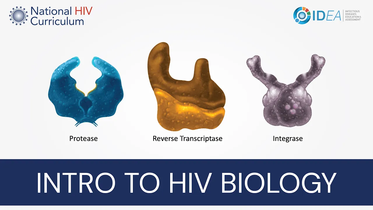
Each of these enzymes plays a critical role in HIV replication. The first enzyme to review is HIV protease. Each virion has approximately 120 HIV protease enzymes, and HIV protease is located outside the viral core.
The HIV protease is a small homodimer consisting of two mirror-image subunits. This enzyme has three functional regions: the flap, core, and terminal domains. The active site for this enzyme is in the center of the two core domains.
The HIV protease acts like a molecular scissors with the opening and closing of the flap region. This opening and closing also enables the substrate to enter and leave the active site of protease. Functionally, the HIV protease cleaves HIV polyproteins at multiple sites, which, after multiple clips, frees the individual HIV proteins.
HIV protease plays a critical role in protein processing of the gag and gag-pol polyproteins during the HIV maturation process very late in the HIV life cycle. The next enzyme to review is reverse transcriptase. Each virion has approximately 100 reverse transcriptase enzymes.
The HIV reverse transcriptase contains two major distinct subunits: the larger p66 subunit and the smaller p51 subunit. The p66 subunit serves more of a functional role with two important enzymatic domains: polymerase and ribonuclease H. The p51 subunit performs more of a structural and supportive role.
The polymerase and ribonuclease H domains each have active enzymatic activity. The p66 subunit is often conceptually shown in a right hand configuration with the fingers, thumb, and palm regions. The major function of HIV reverse transcriptase is to convert HIV RNA into HIV DNA in a highly complex process known as reverse transcription.
The HIV polymerase and ribonuclease H enzyme components play a critical role in this process. Let's look at the third enzyme, integrase. In each virion, there are roughly 100 integrase enzymes.
As noted earlier, some of the integrase enzymes are associated with the HIV RNA genome and HIV RNA nucleocapsid protein as part of the ribonucleoprotein complex. Each of the integrase monomers can self-assemble into dimers, tetramers, and rarely octamers. As shown in this image of the integrase dimer, each integrase protein has three functional domains: the C-terminal domain, the N-terminal domain, and the catalytic core domain.
The catalytic core domain contains a trio of amino acids known as the catalytic triad. The catalytic triad coordinates binding with the divalent metal--usually magnesium--as shown in a simplified two-dimensional rendition in the inset. The bound divalent metal serves as a required cofactor for the enzyme activity.
The catalytic core domain is the active enzymatic region of this enzyme. During viral replication, HIV integrase is bound to HIV DNA, and this complex is referred to as the intasome. In the intasome complex, HIV integrase enzyme provides two key functions: The first is 3-prime processing.
This involves the integrase enzyme cleaving several nucleotides on the 3-prime end of the HIV DNA. The second main function of HIV integrase is strand transfer. This is the process whereby integrase catalyzes the insertion and transfer of HIV DNA into the host DNA.
In addition to producing structural proteins and enzymes, the HIV genome encodes for four accessory proteins and two regulatory proteins. The four HIV accessory proteins are vif, vpr, vpu, and nef. These proteins are not an essential component of the viral structure, and they don t have enzymatic activity, but they act in multiple ways to favor HIV replication and survival.
The first HIV accessory protein to discuss is viral infectivity factor, more commonly referred to as vif. The primary function of this is to counteract the antiviral effect of the human protein APOBEC-3G a host restriction factor that inhibits HIV replication. During HIV reverse transcription, APOBEC-3G can deaminate the nucleotide-based cytosine and convert it to uracil.
Since cytosine normally pairs with guanine and uracil normally pairs with adenine, the action of APOBEC-3G results in multiple guanine-to-adenine mutations in the newly synthesized HIV DNA. This process is known as "G to A" hypermutation. During HIV replication, the HIV accessory protein vif is synthesized, and it clusters near the plasma membrane, which is also where APOBEC-3G is located.
At the membrane, vif binds to APOBEC-3G, and this complex is rapidly degraded by the host cell. The net result is the HIV accessory protein vif has counteracted a host defense mechanism. Viral protein R, or vpr for short, is an HIV accessory protein that is postulated to have multiple roles, including several early in the HIV life cycle.
After HIV enters the host cell, vpr is inside the HIV core. Some models suggest vpr plays a role in modulating the initiation of HIV reverse transcription. Vpr is also thought to facilitate nuclear import of the HIV core.
The next HIV accessory protein to discuss is viral protein U (or vpu), and this protein is not typically present in the mature HIV virion. Vpu has two well-established major roles: First, vpu functions to counteract the host restriction protein tetherin, which is also known as BST-2 or bone marrow stromal cell antigen 2. The human protein tetherin inhibits HIV replication by keeping the budding virion attached to (or tethered to) the host cell membrane.
Before tethering reaches the host cell membrane, it is trafficked internally in the host cell through the endoplasmic reticulum, golgi, and trans-golgi network. This image shows tethering bound to the endoplasmic reticulum membrane. When the viral accessory protein vpu is synthesized, it accumulates at host cell membranes, particularly membranes in the endoplasmic reticulum, as shown here, and in the trans-golgi network.
At this membrane surface, vpu binds to tetherin, initiating several processes that include lysosomal degradation of tetherin and reduced recycling of tetherin. The net result is that vpu reduces tetherin levels at the cell surface, allowing HIV to detach from the host cell membrane. The second major role of vpu is to downregulate expression of the host CD4 receptors on the cell surface.
This occurs due to an interaction between vpu and CD4 molecules inside the cell, predominantly in the endoplasmic reticulum. When vpu binds to the CD4 receptor, this initiates a series of reactions that result in proteasomal degradation of the CD4 molecules. For HIV, the vpu-mediated destruction of CD4 is beneficial since CD4 normally traps the HIV envelope precursor protein gp160 in the endoplasmic reticulum region.
So, with more production of vpu, there s less CD4 remaining in the endoplasmic reticulum, and gp160 can more freely migrate to the cell surface to be part of the newly forming HIV particle. The last of the HIV accessory proteins to discuss is nef. The name, nef, or negative regulatory factor, was originally derived from the mistaken concept that this protein was a negative factor for HIV.
A more recent understanding is that nef enhances HIV replication. Nef has a myristyl group that helps anchor it to membranes. The main role of nef is to downregulate the expression of multiple cellular receptors, including CD4 and CD8 receptors.
Binding of nef to these receptors at the plasma membrane triggers a cascade of reactions that result in the internalization and destruction of these receptors. The modulation of these surface receptors benefits HIV. And finally, let s review the two HIV regulatory proteins tat and rev.
They work to significantly enhance HIV transcription and translation. The HIV protein transactivator of transcription, or tat for short, is a potent regulator of HIV transcription. Secreted tat may also have a role in immune activation.
Inside the host cell nucleus, the proviral DNA (or genomic DNA) can switch between a latent phase, as shown here, to an active phase of transcription that begins after the host cell polymerase binds to the promoter region of the HIV DNA. The HIV regulatory protein tat dramatically increases the efficiency of the viral transcription in the active phase. The last protein to discuss is regulator of expression of virion or rev for short.
The HIV regulatory protein rev is one of the first proteins to be made by HIV. Much of the rev that is synthesized rapidly migrates back into the host cell nucleus. And inside the host cell nucleus, rev shuttles unspliced or incompletely-spliced HIV mRNA from the nucleus into the cytoplasm; thus, the major role of rev is to accelerate HIV protein synthesis.
In summary, this mini-lecture has addressed key aspects related to the biology of HIV, including structure, genome, and the three distinct types of proteins. I hope this review has provided you with some new insights and will inspire you to learn more about HIV. Thank you.
The production of this National HIV Curriculum Mini-Lecture was supported by funding from the Health Resources and Services Administration.