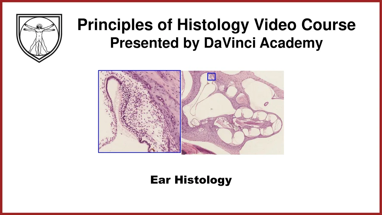
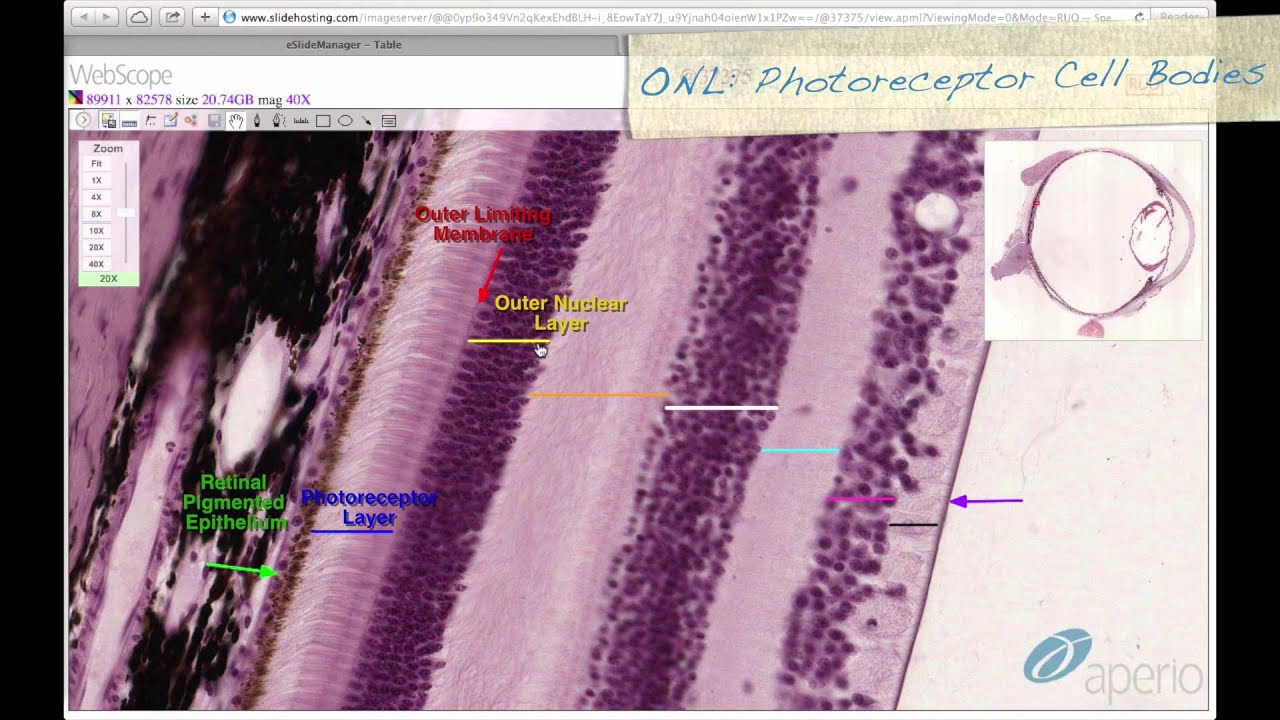
welcome to the da Vinci Academy hystology video course the entire video course is available on YouTube and covers all of the fundamental principles of hystology and relevant cell biology you can find all the videos from the course by clicking the hystology playlist Link in the description below and then you can access the corresponding practice questions and histology Lab videos by going to our website which is also linked in the description below this is the fourth and the last video in the special sensory series and is dedic at to the macroscopic and histological overview of the
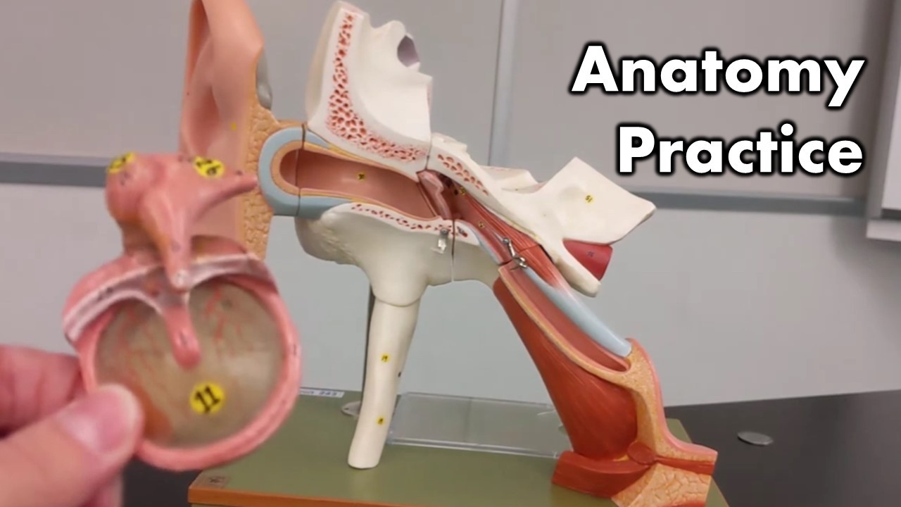
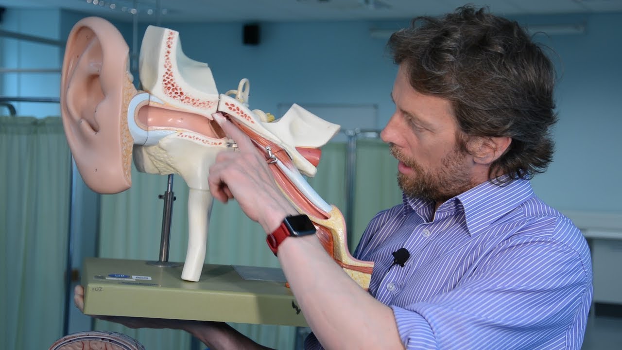
ear we'll begin with the anatomic overview of the ear our ears are subdivided into three segments the external ear which is right here that I'm circling right now is comprised of the Oracle also known as the PA which is this fleshy and protruding structure on either sides of our heads that are specialized to gather sounds which channel the sound waves into the next part of the external ear known as the external auditory meatus this is a narrowed Canal that we can stick our finger into if we so choose the external ear ends at the tanic
membrane this thin tissue paper like membrane which vibrates based on the sound waves that are coming in and it separates the external ear from the next segment of the auditory system and that is the middle ear this middle ear is comprised mostly of air filled chamber and a cavity so the middle ear begins with the tanic membrane it contains these three small bones called the ticles from lateral to medial these ticles are known as the malas Incas and stapes and another important structure of the middle ear is the auditory tube this auditory tube is a
narrow passageway which connects the midar cavity to the nasal fairings this auditory tube is usually collapsed we can open this in order to equalize the air pressure within the middle your cavity with the surrounding environment so when we're taking off in an airplane or climbing up an altitude and we're feeling a little uncomfortable in our heads because of the different pressure within the middle year we can equalize that by opening the auditory tube which is commonly known as popping our ears the third and the most medial as aspect of the ear is the inner ear
and this part is embedded within the Petrus region of our temporal bone the inner ear as you can see is really complex in its Anatomy which we'll look at in a little more detail later but for now just know that the inner ear has the outer bony part and in the inside we have really delicate membranous Labyrinth that follows all the Contours of the Bony casing and this membranous Labyrinth is itself filled with fluid and in its entirety is kind of supported and floating within the fluid of slightly different composition again more on that later
but let's first start with the histology of the external ear the Oracle or the PA is covered on the external side by the skin so we can expect to see some typical skin histology there including hair follicles in the inside of course there's dermal tissues and subdermal tissues like a lot of fatty connective tissue but the structural support of the Oracle and the reason why our oracles maintain its shape for the most part is due to the elastic cartilage core in the inside which is helpful in our ability to bend our Oracle and change its
shape push it against the head but it pops right back to its original shape shape usually that's thanks to the elastic cartilage the external auditory meatus is also covered by skin but here there are some sebaceous glands that are a little more substantial that produce cerumin which is also known as the ear wax so these specialized sebaceous glands of the external auditory meatus are known as the ceruminous glands in terms of the structural support that keeps the external auditory meatus open at all times is thanks to the elastic cartilage laterally but as you can see
here towards the medial aspect the main structural support is provided by the temporal bone moving on to the inner ear we'll start with the macroscopic or anatomic overview of this complex structure the inner year is divided into the Bony outc casing called the Bony Labyrinth which is filled with fluid called Peril lymph this complex anatomy of bony outc casing has several structures these three loopy structures are called the semicircular canals because they look like semicircular tunnels and since they're filled with fluid we'll call them canals instead you may note that these semicircular canals are oriented
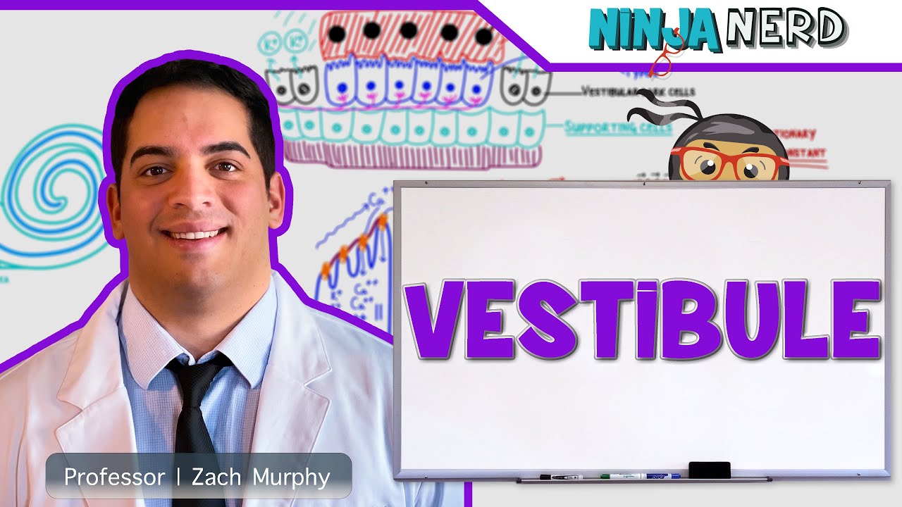
orthogonal to each other in three dimension next we have the vestibule which is this bulbous area right here the space inside side the vestibule is continuous with the space within the semicircular canals so it is still filled with paril lymph and the last part of the Bony Labyrinth is this snail shell-shaped structure called the CA and this snail shell looking structure makes about 2 and 3/4 of a turn and the space inside it is continuous with the vestibule and is filled with the Parry lmph as well you may have noticed this darker gray stuff that
is inside the these various regions of the Bony Labyrinth these network of tubes inside the Bony Labyrinth is what makes the membranous Labyrinth so this so this long and complex tube and bulbous structures is called a membranous labyrinth and it is filled with a different type of fluid called the endolymph the composition of endolymph is set to be quite unique but overall but overall it is similar in its ionic composition to the intracellular fluid whereas the composition of the parm is set to be closer to that of the interstitial fluid since the Bony Labyrinth had
these three sub regions these complex tubular structures of the membranous labyrinth to have their specific Regional names the membranous Labyrinth tube that runs within the semicircular canals are called the semicircular ducts and you may have noticed that each semic cular duct ends in these ular or slightly dilated regions and each one of these dilated region is called the ampula or ampulle in plural so we have the semicircular ducts inside the semicircular canals ducts in the canal is a good way to remember this Arrangement and then these bulbous ends of the semicircular ducts are called the
ampula within the vestibule this large bony casing we have two dilated regions of the membranous labyrinth called the utricle and SACU utricle is closer to the ampulle and SACU is further away from the ampulle but still utricle SACU and semicircular ducts are all continuous channels filled with endoline and finally within the ca we have this continuation of the membranous labyrinth in this coily tube which is known as the clear duct so clear duct is inside the CA and Within These distinct regions within the membranous Labyrinth is where the specialized sensory structures are located within the
ampula of each semicircular duct we have these spots which are indicating the specialized sensory structures called the chiste ulis and these chiste ulis are specialized to sense the angular motion or position of the head the special sense structures of the utricle and SACU indicated by these dark ovals are called the macula the macula within the utricle and SACU are oriented in perpendicular plane to each other and these macula are specialized to sense the linear position or movements of our head in either vertical or horizontal plane and lastly within the clear duct we don't have a
spot but instead we have a really really long linear special sensory structure that goes all the way around following the coil of the clear duct and this linear special sensory structure is called the organ of cordi and it's this organ of cordi that is responsible for the sense of hearing so while the criste ulis and macula of the ampula and utricle and SACU are responsible for sense of balance and motion it is the organ of cordi within the clear duct that is responsible for the sense of hearing and it is for this reason that the
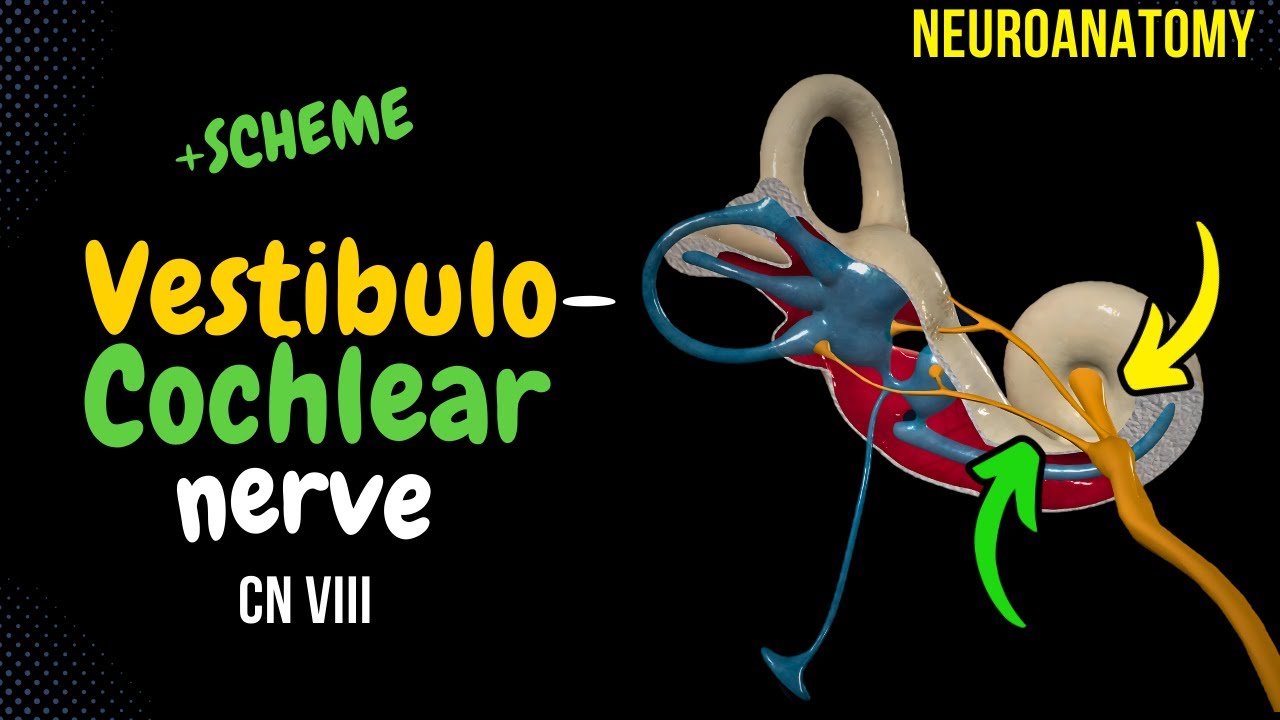
inner year is sometimes subdivided into the vestibular system for the sense of balance and the clear system responsible for the sense of hearing and the cranial nerve that supplies the inner ear is also subdivided into the vestibular branch which carries the Action Potential from from the vestibular system and it also has the clear branch which carries the Action Potential from the organ of cordi of the clear system hence the cranial nerve associated with the inner ear is named the vestibular clear nerve the cranial nerve number eight and here's the actual hystology of the inner ear
and although it's not exact I think approximately this is the plane of section through this complex three-dimensional structure we have to play a little bit of mental gymnastics to figure this out but based on our histological knowledge we can see the Bony Labyrinth forming this bony outc casing of the inner ear and in the inside of the Bony Labyrinth we can see this network of complex tube like structure which is the membranous labant so this is most likely a utricle and the beginning of the semicircular duct and this might be a semicircular duct that we
cut in a cross-section this is the permp filled space outside of the membranous labyrinth which would be filled with endolymph so this region of the Bony Labyrinth would be the vestibule and this might be the beginning of the semicircular Canal likewise here too and over on this side we have the Bony outc casing forming this complex rotating structure or spiraling stru structure so this would be the CA and in the inside we would have this delicate network of endol liil clear duct that has been cut in seral cross-section so these would be the continuous coar
duct that has been sectioned in multiple plane due to the rotating or spiraling nature throughout the ca now let's look at the specialized sensory structures of the inner year we'll start with the Christa Andis we have to remember that there are three ampula one each for the three semicircular dots and each ampula contains the specialized sensory structure plural of Christa ulis is Christa ulis the Christa ulis is comprised of the specialized hair cells with hair which are in reality comprised of stereocilia and the specialized nonmotile cyia called the kyum and this structure is embedded in
a gelatinous substance called the cupula and together the chry ulis is responsible for detecting the rotational position and angular movements of the head at higher magnification of one of these cista ulis we can appreciate the Bony Labyrinth out here and in the inside we have the membranous Labyrinth that encloses the endol lymph filled space on the outside of the membranous labyrinth we would have the Peril lymph filled space and here unfortunately this side of the membranous labyrinth is actually collapsed inward a little bit in reality it should be really ballooning out so this is an
artifactual collapse at any rate we can appreciate that when we trace this membranous Labyrinth the lining epithelium becomes quite thick over here and this is the Christa ulis of the ampula and here these tall cells are the hair cells and although it's not quite as clear you can see the apical surfaces of these cells contain these really fine hair which are again the stereocilia and kinocilia and these hairs are embedded in this really amorphous gelatinous substance called cupula so imagine we are rotating our head and the endol lymph moving around based on the momentum of
this fluid it would push the cupula one way or another thereby causing the bending of these hair and based on the amount of the bending sometimes the action potential May Fire from which the cells May initiate an action potential which is transferred through some of these aeren fibers that are in touch with some of these hair cells next let's look at the hystology of the macula there are two macula in each inner ear one in the utricle and one in the SACU similar to the chista empul macula is comprised of collections of hair cells with
again hair comprised of the kyum and stereocilia which are embedded in a specialized membrane called the oolithic membrane and collectively the macula are responsible for detecting the linear movement of the head in either horizontal or vertical direction we were lucky enough to catch the cross-section through a utricle so let's take a look at this area area in a higher magnification and we can appreciate this region of the membranous labyrinth that is comprised of this tall hair cells and these hair cells once again have lots of these stereocilia and kinocilia that are extending out from the
apical surface and these hairs are embedded in this thin membrane that contain a lot of these heterochromatic crystalline structures that are embedded in this structure which comprises this oolithic membrane this crystallin or calcified structures are called the otolith which makes this membrane slightly heavier once again imagine our head moving from a stationary position and the endol Lim moving in One Direction or another and based on the momentum the oolithic membrane would move against the hair that's embedded within it therefore bending the hair one way or another which can then trick an action potential which is
conducted through these aparent fibers out towards the brain and finally the hology of the CA and the ca duct CA is a part of the Bony Labyrinth that is seashell-shaped and it contains in its inside this long tunnel that coils around about 2 and 3/4 of a turn and in the middle of this bony CA there's a bony core called the modiolus it directly translates to the spokes as in the spokes of a wheel this bony core is where the spiral ganglion is located remember ganglion refers to the neural tissue structure that's comprised of the
neural cell bodies and off of these neural cell bodies we have a collection of exons that are emerging and traveling together which forms a clear branch of the vestibular CIA nerve the cranial nerve number eight as for the coil tunnel it is filled with Peril lymph but in the middle of the tunnel there is that clear duct and this clear duct is actually anchored on either side to the Bony Labyrinth so it's not a really delicate tube that is just floating without any Anchorage and due to these Anchorage points the permp filled space is actually
divided into three compartments the Scala vestibuli that is superiorly positioned and hilot trma is actually at the very tip of the CA and the Scala tempany which is positioned below the kar duct and helot trma is where the Scala vestibuli and Scala tempany become continuous with each other when I uncoil the ca into this long tube what we'll learn is that the ca duct is positioned in the middle and of course the ca duct is filled with endolymph and the Peril lymph fied space between the cier duct and the Bony Labyrinth up here is the
scale of vestibuli and below the ca duct is the Scala Tony and where the Scala vestibuli becomes continuous with Scala Tony this little area is the helot trma so from the middle your cavity we have the oval window right here with the stapes attached so when the sound wave comes in and is changed into Bey mechanical energy this St piece kind of Moves In and Out against this little oval window and this fluid will then move through the Scala vestibuli through the hilot trama and through the Scala Tony and this Force built by the pressure
will be released once again into the middle your cavity through a tiny little membranous window called the round window and within this Clea we have the C clear duct and another name for this C clear duct is Scala media and Scala means a ladder so it's a middle ladder if you will we have to remind ourselves that the clear duct is a part of the membranous labyrinth or membranous tube that is filled with endolymph and it's within this clear duct where the organ of cordi is located which is a specialized sensory structure that can detect
sound waves and translated into an action potential the organ of cordi is located in this particular particular membrane that separates the Scala media from the Scala Tony let's look at the hystology of the CIA duct in a little more detail by zooming in onto the boxed area this is just one section of the coiling clear duct within the CA and at higher magnification we can see that this is the Scala vestibuli the per lymph filled space above the colear duct and this thin membrane that separates the clear duct from the Scala vestibuli is called the
vestibular membrane then we have the ca duct also called the Scala media hosting this specialized structure called the organ of cordi on the bottom membrane which is known as the baslar membrane so this basar membrane not only has the organ of cordi but also separates the Scala media from the Scala Tony and in terms of the organ of cordi we can appreciate that this specialized structure is comprised of these specialized sensory cells with kinocilia and stereocilia that are butting off of the inner group of cells and outer group of cells these cells are called the
inner and outer hair cells respectively and their hair are embedded in this non cellular membrane or structure called a tectorial membrane so when the hair bends against the tectorial membrane that's when the action potential is generated which is then conducted through this neuronal fibers and of course the neuron cell bodies are all collected over here as a spiral ganglion and then their distal fibers or axons are all emerging out conducting that action potential to the brain and the space below the basar membrane is a Scala Tony so if I may uncoil this CA and CA
duct one more time we would see something like this again with the oval window here with st AP is attached and vibrating against the Scala vestibuli that's filled with Peril lymph and that sound wave in the fluid would travel through go through the hilot trma then through the scale of tempany before that pressure is released through the round window and within the Scala Media or the ca duct we have the organ of cordi that is all along the basar membrane with their hair embedded in the tectorial membrane and depending on the frequency of the sound
that is being conducted through the OES and into the scale of vestibuli and tempany a specific region of the Scala media particularly the organ of cordi will move just the right amount to bend the hair against the tectorial membrane which will then trigger the action potential so that we register that sound at the accurate frequency or the pitch what's interesting about this long coily tube gr is that the hair cells closer to the oval and round window happen to detect the higher frequency sound or higher pitch sound and those that are closer to the helot
tra register more lower frequency sound so those sounds that are a little more at the base in Pitch thank you for watching this video from The Da Vinci Academy histology video course which is completely available on YouTube to access the corresponding practice questions and histology Lab videos go to our website using the link in the description below [Music] oh [Music]
![Eye Anatomy and Histology of the Outer Layers of the Eye [Special Senses Histology Part 1 of 4]](https://img.youtube.com/vi/u1fUUr1A34Y/maxresdefault.jpg)
![Kidney Anatomy and Histology [Urinary Histology Part 1 of 4]](https://img.youtube.com/vi/Qm4y2fa-kcM/maxresdefault.jpg)
![Histology of the Testes [Male Reproductive Histology Part 1 of 4]](https://img.youtube.com/vi/DXc-L1SfGBU/maxresdefault.jpg)
![Histology of Eye: Lens, and Mechanism of Accommodation [Special Senses Histology Part 2 of 4]](https://img.youtube.com/vi/_L255O4oefs/maxresdefault.jpg)
![Histology of the Ovary and Ovarian Follicles [Female Reproductive Histology Part 1 of 2]](https://img.youtube.com/vi/cDs6goPOD1I/maxresdefault.jpg)
![Histology of the Retina [Special Senses Histology Part 3 of 4]](https://img.youtube.com/vi/zG4ZhMusF4Y/maxresdefault.jpg)
![Liver, Pancreas, Gallbladder Histology [GI Histology 4 of 4]](https://img.youtube.com/vi/_kA9x9FcjWA/maxresdefault.jpg)
![Bone Anatomy, Bone Cells, & Hormonal Regulation of Bone Calcium [Bone Histology Part 1 of 3]](https://img.youtube.com/vi/0N6tsVFOxo4/maxresdefault.jpg)