Hi, how are you? This video is being sponsored by Politec Recife. And if you are from Recife or nearby regions and are looking to take a course in radiology, nursing, clinical analysis, administration or accounting, this place is for you.
Politec, reference in technical education. So, Radiologando staff. Are you okay?
We are here for another video on the channel. And today I'm going to talk about a very cool content, which is about nuclear medicine. Which I particularly love.
And we already have one here that I commented on this subject here. So if you want take a look at the description here or at the description link which I'm sure you'll like too. And today I'm going to give you some introductory aspects, tell you what nuclear mention is, what are its characteristics and how exams are performed there.
But calm down, before starting the video, I would like you there to leave your like and like the video to help our work. And if you are new to the channel, subscribe to the channel and enable all notifications. That way, you stay on top of all the content that is posted here weekly.
And also follow us on social networks, both on @radiologando and @ygorjulioprof so that you can Radiolog closely with us. Without further ado. Let's go to the video?
But before starting the video, I wanted to give you something new . It's a new resource that we at Radiologando have opened for you to increase and raise your level of studies, which is the ward to be a member, in which you can count on plans, which will help you to learn more and more about radiology. So, if you are curious and want to know a little more, click below, in the wing, become a member and watch the video to learn more about this new feature.
Well, as I said, today I'm going to bring you a video that's different from what you 're used to, because there are many people who haven't completed the course yet, who are still studying and have never heard of nuclear medicine. Nuclear medicine, in turn, is one of the specialties that, in turn, also offers diagnostic and therapeutic exams in radiology. Yes, in nuclear medicine, in addition to diagnostic tests, that is, to generate results, we are also able to carry out therapeutic procedures, procedures that are one of the characteristics attributed and linked to nuclear medicine.
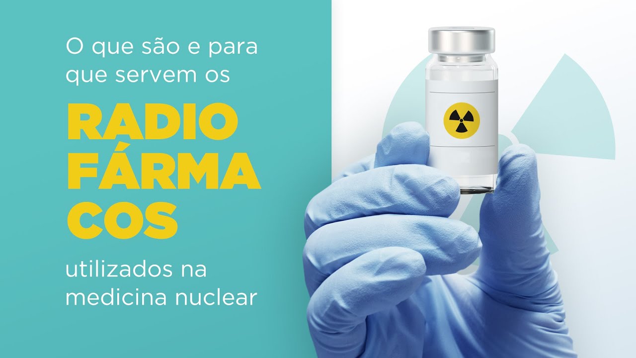
Also different from most radiognostics, many people think that in nuclear medicine, radiological contrast is applied , but actually what are used in nuclear medicine are radiopharmaceuticals, which in turn is a radioactive chemical compound , which in turn will having an affinity with some organ or system of the human body that will also emit radiation in order to form an image. And speaking of image formation, in nuclear medicine it happens totally differently too. In common radiodiagnosis, the equipment will basically emit the radiation and the equipment itself will absorb this radiation in order to form the image.
In nuclear medicine, however, it is the opposite. The patient is going to emit radiation and the machine, in turn, will have the simple function of absorbing this energy, capping this energy, and then forming the image. The term used in nuclear medicine to be able to perform the exams would be called scintigraphy due to the physical phenomenon that occurs in the capture process and the entire process of energy processing for image formation.
So there are several types of exams to be performed. Bone scintigraphy, pulmonary scintigraphy, myocardial scintigraphy , among many others. Not to mention also that nuclear medicine has two, remembering that nuclear medicine is not a diagnostic technique, it is a medical specialty and in this medical specialty there are two imaging diagnosis techniques which is aeaa is single emission computed tomography, it is emission tomography of deposits.
Differently from what is used in the vast majority of radiognostics, which are X-rays, in mecinuclear gamma rays are used or even in the technique in which a different process for the formation of the image will occur . However, regardless of the type of drug or even a type of rays that is used in nuclear medicine, they will act in a specific way . After all, nuclear medicine imaging is more physiology-oriented than anatomy-oriented.
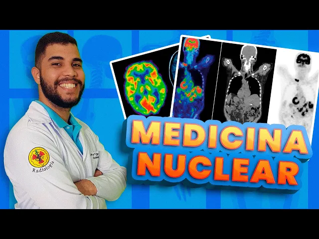
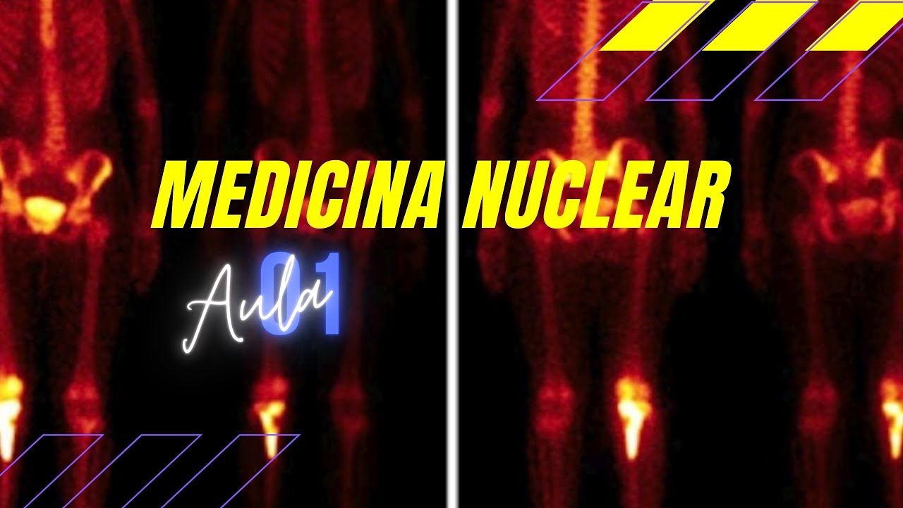
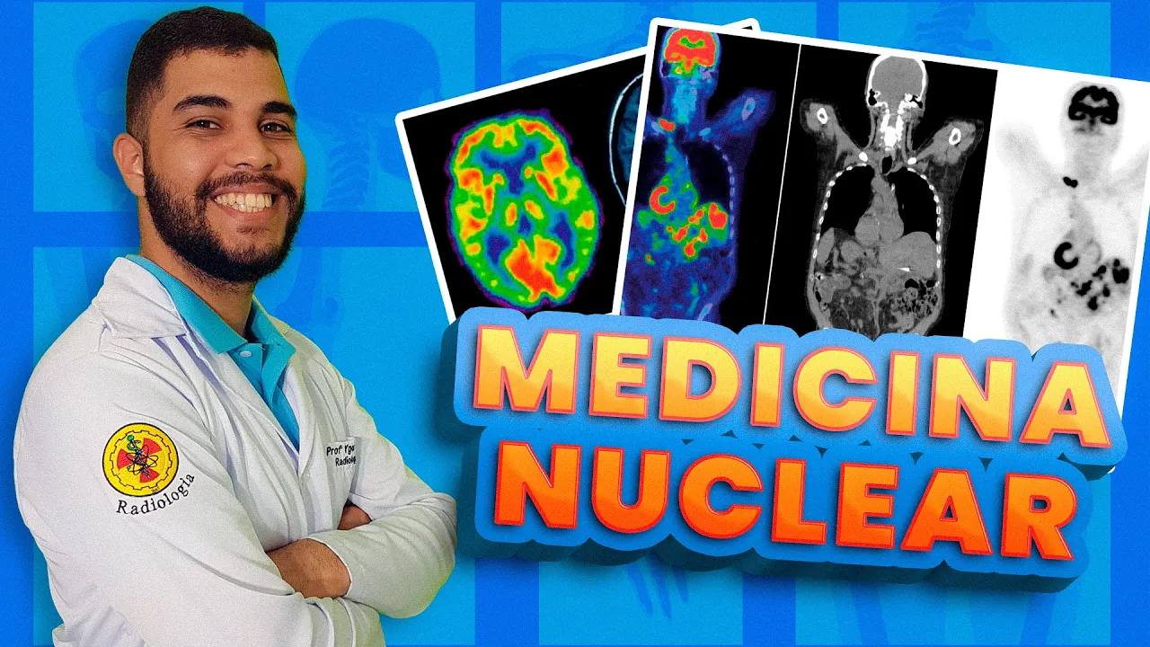
You are now seeing images of scintigraphy on your screen , that is, images that are performed in nuclear medicine, both technically and technically, and you can see that the image is not anatomical at all, why? The image is more physiological, it aims to demonstrate how that organ or even that system is working and if the patient has any metabolic alteration, the image will demonstrate this, not to mention that in the nuclear mess, each organ for in turn, it will react differently to each type of radiopharmaceutical that is used, so several different types of radiopharmaceuticals are used and all of them with a purpose to demonstrate possible metabolic changes or not in relation to the human body, so I cannot put a radiopharmaceutical which is used for a bone scintigraphy the same for myocardial scintigraphy or vice versa. For each organ for each system there is a specific radiopharmaceutical to be used.
Remembering that the radiopharmaceutical is a chemical compound plus a radioactive compound that are joined together that are marked to be administered to the patient and different from the common technique when the patient receives the radiopharmaceutical, why can't he immediately perform the procedure? This radiopharmaceutical has to travel throughout the body until it reaches the organ or system of interest and for this there is a biodistribution time for each drug, sometimes the patient to be able to enter the examination room to be able to perform the procedure may take two, three and even four hours as in the example of static renal scintigraphy, so it is very important for us to know all this. Of course, we learn this gradually, that is, little by little.
And you must be asking yourself , okay, professor, as a technician or as a technologist in radiology, can I work in nuclear medicine? Yes, both technicians and technologists can work in nuclear and detail, it 's not that you necessarily need to have a qualification course , have an extension course, have a postgraduate degree or a specialization to work. Of course, this helps, not only on your resume, but also on your knowledge.
But you can work in nuclear medicine , you can start to learn to work in nuclear medicine, you can do an internship in nuclear medicine so that you can learn how the entire procedure is performed . But know that it's totally different. Radiodiagnosis.
One of the very important requirements that is applied to nuclear medicine is radiological protection . As I said, it is not the equipment that will emit radiation, it is the patient that will emit radiation after receiving the administration of the radiopharmaceutical. So basically, the professional who works in nuclear medicine must use the lead apron, the thyroid protector and the eyeglasses with blisters, why?
It's not just what will emit radiation. In the nuclear medicine service there is a radiation called background radiation or radiation which in turn is very characteristic of the hot sole and also of the procedure rooms where patients undergo examinations. So it is very important for us professionals to use all PPE throughout the working hours.
In order to optimize, reduce the amount of radiation absorbed during our work. Another characteristic that can occur in nuclear medicine is contamination. Unlike other radiognostic services , we simply have irradiation there, but in nuclear medicine, in addition to irradiation, we also have contamination.
Of course, it is not that we are going to have contamination, but that there may be contamination, but the contamination, in turn, is controlled. Don't worry, you won't be at risk and there won't be any kind of accident. Rest assured.
These are just criteria that can happen in the nuclear service. And if you want, I can bring a video here explaining the difference between irradiation and contamination. The difference between each of them and their characteristics.
But yes, contamination can occur while performing a procedure. After all, the radiopharmaceutical will be inside the patient. Yes, it depends a lot on the clinical condition he is in, he can vomit, he can urinate and in this vomit this urine can contain radioactive material, so that area is contaminated, but it is not contaminated for the rest of his life, even because the radiopharmaceutical is the radioactive material it has a physical half-life, which is another criterion, another characteristic, that is, the half-life time will basically determine the final energy of this radioactive material .
There are several types of time in nuclear medicine , in the technique the most used is the ninety-nine stable target, which in turn has a half-life there of approximately six hours. It means that every six hours your energy will drop by half, that is, in three, four days at most that area that was contaminated, that area is now totally free of contamination and totally safe. So there's no way to worry.
Not to mention also that in the radiosoxide technique most used, which is fluor eighteen, it has a half-life of approximately two hours. So, note that the half-life attributed to radioactive compounds that are used for radiopharmaceutical labeling has a short half-life and this greatly favors radiological protection and biosafety for everyone involved, both patients and professionals. And to finish the video, we also have the nomenclature that is assigned to the exams that are carried out in the nuclear service.
Unlike what we see in the radiodiagnostic image as radiopaque, radiolucent, hyperattenuating, hypoattenuating, in nuclear medicine, how we work with capture, in the image when we have a hot area , that is, an area that has a much higher concentration of radiopharma , we said that in that area there was a hyper-uptake and in the cooler areas, in the areas that are a little darker, we call that area a low radiopharmaceutical. So, that area has a hypocapitating feature. Hypercapitative is very uplifting .
Hippocapitante has little uptake and where we do not have any signal it is characterized as an absence of signal. So it is very common also in scintigraphic images that we use the terms that region is very hot. This region is very cold.
It actually means that this region has a high concentration of radiopharmaceutical and that region has a low concentration of radiopharmaceutical. So, did you learn? I hope so.
I also hope you enjoyed the video, but if you still have any questions, leave it here in the comments and Radiologando will answer for you. But if you don't have any doubts either, leave it here in the comments that I want to know, if you want more new topics like this. And also tell me what you thought of this video and if it helped you in any way.
Don't forget to like the video, subscribe to the channel and activate all notifications to stay on top of all the content and also follow us on social networks, both at radiologando and at at sign so you can radiologan up close with us. I'm sticking around here and I'll see you in the next video. Thanks.